1961
Notes upon the intercostal arteries in some snakes
Publication
Publication
Bijdragen tot de dierkunde , Volume 31 - Issue 1 p. 53- 57
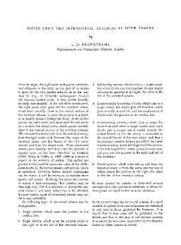
From its origin, the right aortic arch passes anteriorly, and obliquely to the right; in this part of its course it gives off the two carotid arteries, or as the case may be (e.g., in Uropeltis melanogaster (GRAY)) the common carotid trunk. It then curves dorsally, medially, and caudally. At the end of the dorsal curve, the right aortic arch gives off the vertebral artery, which runs cranially, close to the ventral surface of the vertebral column, to enter the parietes at a greater or smaller distance behind the head. In its further course, the right aortic arch fuses with the left aortic arch to form the dorsal aorta, which passes caudally close to the ventral surface of the vertebral column. The intercostal arteries arise from the vertebral artery, from the right aortic arch (between the origin of the vertebral artery and the fusion of the two aortic arches), and from the dorsal aorta. These intercostal arteries pass dorsally, and they enter the parietes in varying ways, as has been described by BEDDARD (1903; 1904a, b; 1906a, b; 1908; 1909) in a series of papers on the anatomy of snakes. This author has pointed to the possible taxonomic value of the differences shown by the various genera and species, which he examined. However, before definite conclusions can be drawn, it will be necessary to examine more genera and species. Studying the intercostal arteries of snakes is time-consuming; their number may be very high (e.g., 156 in a specimen of Xenopeltis unicolor Reinw.), and every artery has to be checked, because various types of intercostal arteries may occur in one individual. The following types of intercostal arteries can be distinguished.
| Additional Metadata | |
|---|---|
| Bijdragen tot de dierkunde | |
| Released under the CC-BY 4.0 ("Attribution") License | |
| Organisation | Naturalis journals & series |
|
Brongersma, L. D. (1961). Notes upon the intercostal arteries in some snakes. Bijdragen tot de dierkunde, 31(1), 53–57. |
|