1975
Light and electron microscopic studies of the ascus top in Sarcoscypha coccicea
Publication
Publication
Persoonia - Molecular Phylogeny and Evolution of Fungi , Volume 8 - Issue 3 p. 259- 271
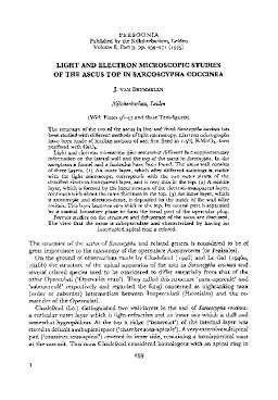
The structure of the top of the ascus in live and fixed Sarcoscypha coccinea has been studied with different methods of light microscopy. Electron micrographs have been made of median sections of asci first fixed in 1.5% KMnO4, then postfixed with OSO4. Light and electron microscopy give somewhat different but supplementary information on the lateral wall and the top of the ascus in Sarcoscypha. In the ascoplasm a funnel and a funiculus have been found. The ascus wall consists of three layers. (1) An outer layer, which after different stainings is visible with the light microscope, corresponds with the two outer strata of the stratified electron-transparent layer, and is very thin in the top. (2) A middle layer, which is formed by the inner stratum of the electron-transparent layer, continues with about the same thickness in the top. (3) An inner layer, which is anisotropic and electron-dense, is deposited on the inside of the wall after meiosis. This layer becomes very thick in the top. Its central part is separated by a conical boundary plane to form the basal part of the opercular plug. Former studies on the structure and dehiscence of the ascus are discussed. The view that the ascus is suboperculate and characterized by having an interrupted apical ring is refuted.
| Additional Metadata | |
|---|---|
| Persoonia - Molecular Phylogeny and Evolution of Fungi | |
| Released under the CC-BY 4.0 ("Attribution") License | |
| Organisation | Naturalis journals & series |
|
van Brummelen, J. (1975). Light and electron microscopic studies of the ascus top in Sarcoscypha coccicea. Persoonia - Molecular Phylogeny and Evolution of Fungi, 8(3), 259–271. |
|